脱毛について
脱毛ができない場合もありますか?
●妊娠中もしくは妊娠の可能性のある方
●重度のケロイド体質、重度のアトピー体質、重度の皮膚疾患のある方
●光アレルギーのある方、光が禁忌となるお薬を服用中の方、てんかんの既往のある方
●処置の部位にタトゥー・感染がある方痛みにはとっても弱いです。本当に痛みなく施術できるのでしょうか?
従来のショット式は強いレーザー量を一瞬で照射していたため痛みを強く感じることが多い施術として知られています。
蓄熱式脱毛は、毛根周囲の温度を徐々に上昇させていくことで、強い痛みを感じることはあまりありません。
ただし痛みの程度は個人差が大きく、表面麻酔を併用するなどして痛みを軽減することも可能ですので、脱毛カウンセリング時にお申し出ください。長い時間施術をするのは大変です。1回の脱毛時間はどれくらいでしょうか?
部位やエリアの大きさによって異なりますが、片脇で1分程度です。
最新の蓄熱式脱毛機器であるMotus Axは、術中冷却を行っていることから術後の冷却が原則不要となり、実質施術時間が大幅に短くなります。男性です。Vラインは男性でも形を希望することはできますか?
男性のVラインでも形を整える「デザイン脱毛」はもちろん可能です。 詳しくは「お問い合わせ」またはTELにてお気軽にご相談下さい。

Vライン脱毛をしたいと思います。全てなくなってしまうのでしょうか。
Vラインはデザイン脱毛が可能です。
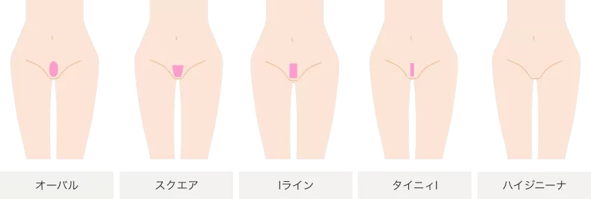
介護脱毛であれば、「ハイジニーナ」という全脱毛がおすすめです。
「完全になくなるのは少し恥ずかしい」という方もおられるかもしれません。
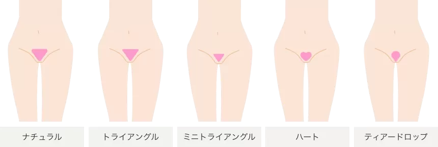
その方には下の図のような「デザイン脱毛」をおすすめします。
当院では「介護脱毛」も多くハイジニーナを選択されるも多いのですが、20-30代の方では「ミニトライアングル」「トライアングル」を選択される方が多いです。

医療脱毛何回も通わないといけないのですか?
医療脱毛は脱毛サロンと違い、高出力レーザー機器を使用できますので概ね3-5回でほぼ満足のいく効果を得られる場合がほとんどです。
ただし、皮膚の状態・毛質・毛量・施術直前の処理状態によって異なります。
というのも、毛の状態によって毛根・毛周囲に吸収されるエネルギー量が異なってしまうからです。
お客様の皮膚状態を確認することで、より正確な情報をお伝えすることができます。
詳しくは脱毛カウンセラー・医師の問診時にお尋ね下さい。何度も通うのは大変です。どれくらい通えば良いのでしょうか?
通常、5回を基準に施術いたします。
しかし、毛には生えるサイクルがあり、減耗効果が最も得られる毛の成長期で施術を行なっても1回の施術で脱毛できるのは全体の20%程度とされています。
また、部位により毛の生えるサイクルが異なりますので、施術後に適切な時期をご案内いたしますが、5回の施術でおおよそ1年となります。エステの脱毛とどこが違うのでしょうか?
当院では医療用脱毛レーザー機器を使用します。
脱毛サロンは光脱毛がメインとなり、毛が薄くなることで減毛効果があたかもあるかのように錯覚しますが長期減毛効果はそもそもありません。
エステ脱毛に比べ医療脱毛では毛の幹を破壊してしまうため比較的少ない回数で長期減毛できることが最大の違いとなります。
注意事項について
中途解約手数料について
中途解約の場合は、一般的に手数料が発生します。手数料は、以下の2つの金額のうち低い方の金額となります。
5万円
契約残額の20%に該当する金額
このように、どんなに高くても5万円となるため、法外な手数料を取られる心配はありません。
契約残額の計算は、以下の例を参考にしてください。
例)全身脱毛5回30万円コースを2回で中途解約する場合
1.脱毛1回あたりの金額を計算(300,000÷5=60,000円)
2.施術を受けた2回分の金額を計算(60,000×2=120,000円)
3.契約残額を計算(300,000-120,000=180,000円)
契約残額は180,000円となります。
この場合、解約手数料は36,000円となりますので、180,000円から解約手数料36,000円を引いた144,000円が返金されることになります。クーリングオフについて
1.お客様は、契約書面を受領した日から起算して8日間以内であれば、関連商品を含め、書面により契約を解除することができます。(これを「クーリング・オフ」といいます)。クーリング・オフをした際に、当院が、契約の解除に伴う損害賠償又は違約金の支払いを求めることはありません。又、提供済みの役務の対価その他金銭の支払いを求めることはありません。又、当院が契約に関してお客様から金銭を受領しているときは、速やかに全額返金いたします。
2.お客様が当院より事実と異なることを告げられそれを事実であると誤認したことにより、又はお客様が当院より威迫され困惑したことにより、クーリング・オフを妨害された場合、お客様は当院から改めてクーリング・オフができる旨を記載した書面を受領し、当院から説明を受けた日から起算して8日間以内であれば書面によるクーリング・オフをすることができます。
3.クーリング・オフは、お客様がクーリング・オフ書面を当院宛に発信した時に、その効力が生じます。
4.クレジット等をご利用の場合の清算は、各クレジット会社所定の方法によりますので、詳しくは各クレジット会社の規約等でご確認ください。
<<書面送付先>>
契約された各院宛に送付をお願いいたします。※お電話での通知は受け付けておりません。必ず書面を郵送いただけますようお願い申し上げます。ご解約について
・引っ越し・転勤等によるコース途中での解約が可能です。コース未消化分の残金は返金いたします。 また、契約から8日以内であれば、クーリングオフによる全額返金も可能ですので、詳しくはクリニックまでお問合せください。
※中途解約時には所定の手数料を頂戴しております。
※解約ができる期間は2年以内までとさせていただきます。お支払い方法
下記のいずれかからお支払方法をお選びいただけます。
現金
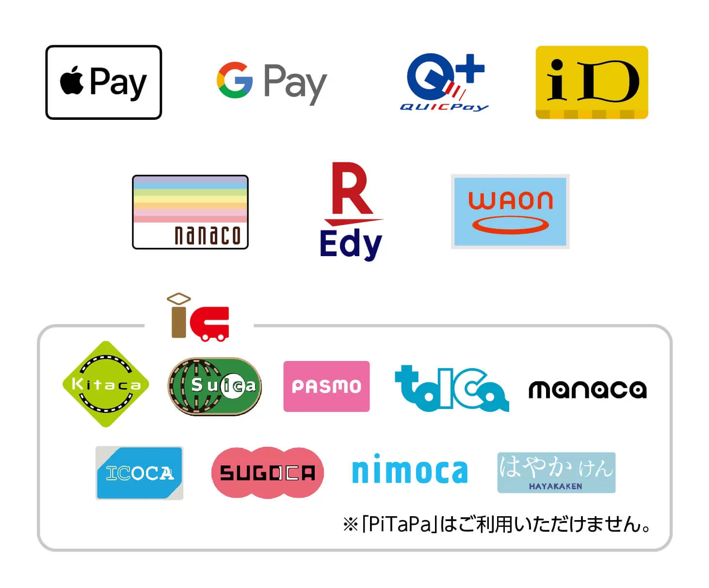
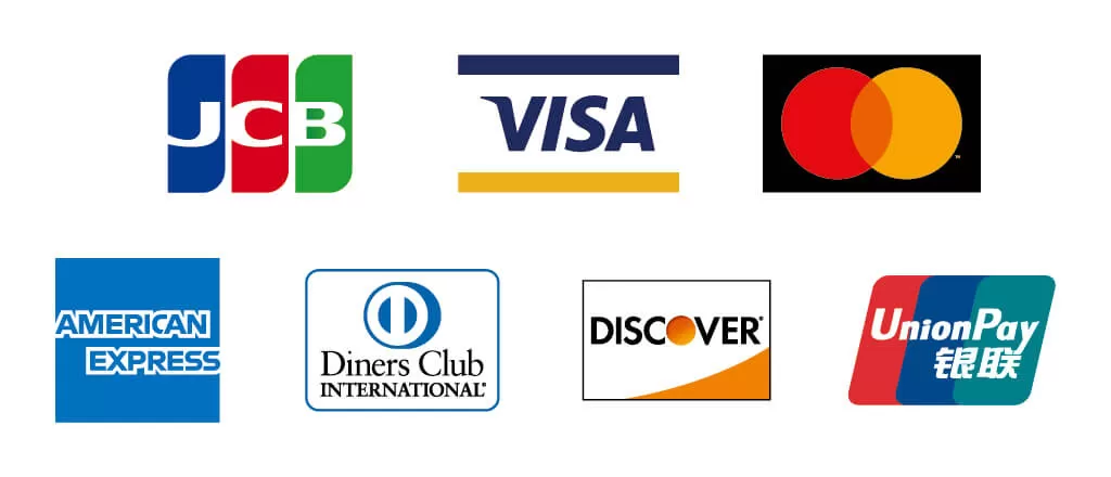
クレジットカード・電子マネー
<下記の支払い方法が使用できます>


月額プラン 月額プランとは、施術合計金額を分割でお支払いいただく医療機関専用のローンです。月々の支払が3,000円から始めることができます。支払回数についてはカウンセリングの際にお気軽にご相談下さい。
未成年の方は必ずご確認ください。
未成年の方は代表親権者様の同意書及び代表親権者様の身分証のコピーの提示
コピーの提出が必要となります。
13歳未満の方のご契約はお断りさせていただいております。
高校生以下の方は親権者様の同伴が必要です。
親権者同意書の内容確認のため、親権者様にご連絡させていただく場合がございますのでご了承ください。
親権同意書のダウンロード
ホワイトニングについて
目立った効果が期待できないケースは?
ホワイトニングは沈着した色素を分解して、もともとの歯の色に近づけるものです。従って、加齢によって変色している歯や、神経がなく黒ずんでしまった歯、テトラサイクリン歯(以前使用されていた、テトラサイクリン系の抗生物質の副作用による灰色の歯)などには、効果が期待できません。また、もともと歯の表面に細かい傷がある場合は、表面の光の反射が原因で、白さを実感しにくくなります。
詰め物や被せ物にもホワイトニングできるの?
ホワイトニングの薬剤で白くできるのは、基本的には天然の歯だけとなります。従って、ホワイトニングをすることによって、詰め物や被せ物との色が合わなくなるケースもあります。詰めた時期が早ければ、それほど目立たない場合もあります。当院では歯科医師が事前にチェックしご説明いたします。
後戻りって何?
前述した通り、ホワイトニングの効果には持続期限があります。食事などによって、再び歯が黄ばんできます。これを、後戻りといいます。ステインが付着すると、後戻りが速まりますが、ホームホワイトニングを併用すれば、後戻りを防げます。
効果はどのくらい持続するの?
一般的には、オフィスホワイトニングで3ヶ月から6ヶ月程度となります。ホームホワイトニングでは半年から1年くらいが目安となります。ホームホワイトニングは、時間をかけてホワイトニングするので、薬剤を十分に浸透させるので白さが長持ちします。
ホワイトニングはどのくらい白くできるの?
歯の白さの度合いを見る目安として、シェードガイドというものがあります。白い順に、16段階に分けて色の違いを示したものです。オフィスホワイトニングでは、今より2段階ほど白くなります。じっくりと時間をかけて、より白くできるホームホワイトニングでは、現在の色から、およそ1から4段階くらい白くすることができます。ただし、白さをアップするのにかかる時間は人それぞれです。また、エナメル質に沈着した色素を分解するものなので、自分の歯のもともとの白さを超えることはできません。
オフィスホワイトニングとホームホワイトニングはどちらが白くなる?
白さを実感できるスピードで比較するなら、オフィスホワイトニングの方がお勧めです。高濃度のホワイトニング薬剤を扱い、光などを照射して、その反応を高める集中的な施術ができるからです。しかし、ホームホワイトニングは低濃度の薬剤であっても、毎日2時間、2週間程度継続し、じっくりと薬剤を浸透させることができます。ホームホワイトニングはご自身で白さのコントロールができます。
ホームホワイトニングとセルフホワイトニングは同じもの?
違います。歯科医師のいないサロンなどに足を運んで、自分で行うのがセルフホワイトニングです。歯科医師がいないので、オフィスホワイトニングで使われる医薬品のホワイトニング剤を扱うことはできません。従って、漂白するホワイトニングではなく、主にポリリン酸やメタリン酸など市販されている薬剤を使った、黄ばみやステインの除去となります。歯科医師は歯の漂白ができる過酸化水素という薬剤を使用できます。
オフィスホワイトニングとホームホワイトニングの違いは?
歯医者さんで施術を行うのがオフィスホワイトニングで、歯医者さんの指導のもと、提供された薬剤やマウストレーを使って、自分で行うのがホームホワイトニングです。どちらも、過酸化水素を成分にした薬剤で漂白を行うことは同じです。また、オフィスホワイトニングでは、高濃度の過酸化水素を扱った、短時間の施術(1時間程度)となります。一方、ホームホワイトニングは低濃度の過酸化水素の薬剤を使い、ご自身でマウスピースを用いて、時間をかけて白くしていきます(1日2時間で最低でも2週間程度)。
市販のホワイトニング歯磨き粉とは作用が違うの?
白くする成分が違うのでその作用も違います。歯医者さんで行うホワイトニング剤は、主に過酸化水素(オキシドールの成分)が含まれる薬剤を使うことで、歯を漂白しています。一方、日本で市販されているホワイトニング歯磨き粉は、薬事法の規制があるため、過酸化水素を成分としたものはありません。歯を白くするための、主な成分はポリリン酸ナトリウムやポリエチレングリコールなどで、ステインを除去するためのもので、漂白する効果がありません。
クリーニングとホワイトニングは何が違うの?
クリーニングでは、主に自分では落とせない隅々の歯垢や歯の表面にこびりついたステイン(着色汚れ)を機械で取り除きます。一方、ホワイトニングは歯に染み込んでいる色素を分解して、漂白します。主に、歯の表面にあるエナメル質や象牙質の表面を漂白することで、歯の自然な透明感や明るさを増すことができるのです。
あたまのかたち外来について
頭蓋変形が最近注目されてきたのには何か理由はありますか?
1992年にAAP(米国小児科学会)が「Back to Sleep(仰向け寝)」キャンペーンを大々的に行いました。これは乳児を仰臥位(supine position)で寝かせることで、社会問題になっていたSIDS(乳幼児突然死症候群)を減少させることが目的でした1)。このキャンペーンのおかげで、1992年から2000年にかけて死亡率の減少が得られましたが、その反面後頭部の扁平化が問題化してきたのです2)。位置的頭蓋変形は1992年以来4倍の発生率が報告されるようになりました。アメリカでは60人の赤ちゃんのうち1人に頭蓋変形が認められると報告されています。
1. American Academy of Pediatrics AAP Task Force on Infant Positioning and SIDS: Positioning and SIDS. Pediatrics 1992;89(6 Pt 1):1120-6.
2. Diagnosis and Management of Positional Head Deformity, Wendy S.Biggs, M.D.,University of Michigan Medical School, Ann Arbor, Michigan; American Family Physician, VOLUME 67, NUMBER 9 / MAY 1, 2003頭蓋変形の頻度はどれぐらいなのですか?
乳児期の早期にはおおよそ40%前後の赤ちゃんに認められることが報告されていますが、頭蓋変形の統一した定義がないため、報告は論文により大きく異なっているのが現状です。
頭蓋変形は英語でなんというのでしょうか?
頭蓋変形の種類によって名称が異なります。◎Positional skull deformity◎Positional head deformity◎Oblique head◎Plagiocephaly等、様々な名称で呼ばれています。Misshapen headという一般的な名称もよく使用されます。『頭蓋変形』の原因には様々な疾患群が含まれており、そのために名称も色々多用されているのが現状です。
頭蓋変形の定義を教えてください
広く使用されている『頭蓋変形』という言葉ですが、統一した定義は現時点ではありません。海外では「Baby misshapen head」という一般的な名称がありますが、そこに含まれる疾患群は様々です。
あたまの骨は数種類の骨の組み合わせだと聞きました。詳しく教えてください
乳児期の頭蓋(ずがい)は八個の骨格が脳や延髄を囲む円蓋(えんがい)を形成します。成人の場合、頭蓋の骨格はすべて縫合(ほうごう)と呼ばれるほぼ可動のない接合(せつごう)によってお互いが連結されていますが、乳児の場合は線維性縫合(せんいせいほうごう)は形成しますが、癒合(ゆごう)はしていません。この癒合が行われるのは生後およそ2歳から3歳頃です。
骨が癒合していないことで、急激な脳の成長に対応しています。脳の成長は胎児期から始まり、出生時の脳重量は約300gとなります。その後、生後6カ月で約2倍、2歳では約3倍になります。4歳から6歳で成人の脳重量(1,200〜1,500g)の約95%に近づくことが知られています。
Tokyo Baby’s Head Clinicについて
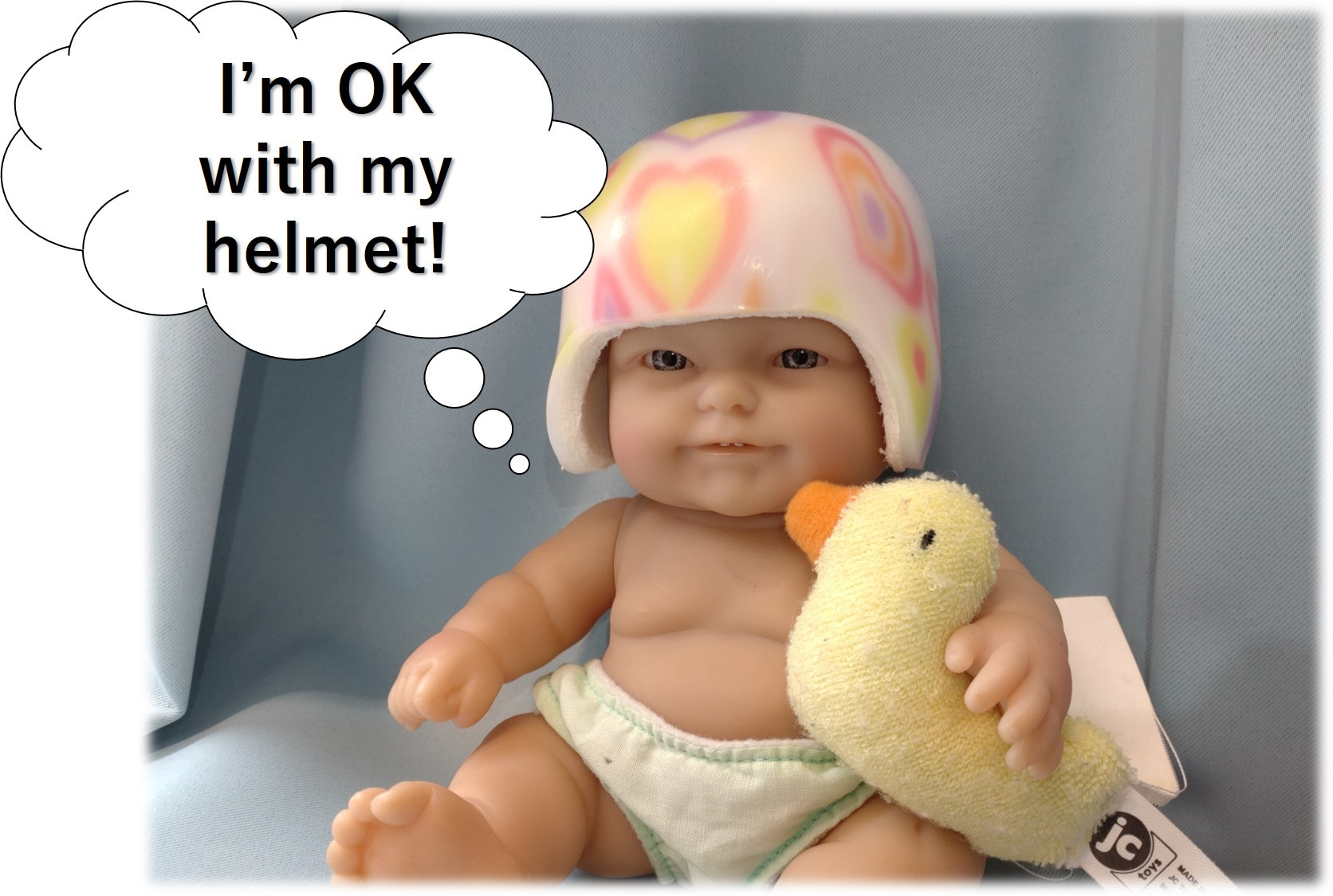
I feel that the hurdle to visit the hospital is high, and I am too busy to visit on weekdays due to balancing work and childcare.
Mitaka Hiro Clinic, on Saturday, Sunday, and holidays are open to the customers with a full reservation system. If you are worried about your baby’s head shape, feel free to just talk to our specialist doctor without any hesitation.
What exactly does a doctor do during the examination? I’m worried that the baby might feel pain or discomfort.
The head shape specialty clinic is staffed by experienced pediatric neurosurgeons. The examination and diagnosis are performed with the family present to ensure that the baby is not anxious or uncomfortable. In some cases, a medical net with excellent stretchability may be used to observe the shape of the skull more closely. The main items observed are the degree of patency of the anterior and posterior fontanelles, the position of the ears, the degree of frontal protrusion, and mandibular displacement, among others. In addition, we will ask about birth weight, events during pregnancy, and changes in head circumference, so please bring your “Maternal and Child Health Handbook” to the examination.
I heard that there is a “natural correction” for cranial deformation. Can it sometimes heal on its own without treatment?
For positional plagiocephaly, the severity of the condition is first classified. If it is mild to moderate, natural correction can be expected to some extent, but if it is severe to extremely severe, natural correction is extremely difficult. It is important to discuss with a specialist physician, taking into account the age of the child and the applicability of helmet correction therapy.

When is it possible to correct cranial deformities? What is the appropriate age range for treatment?
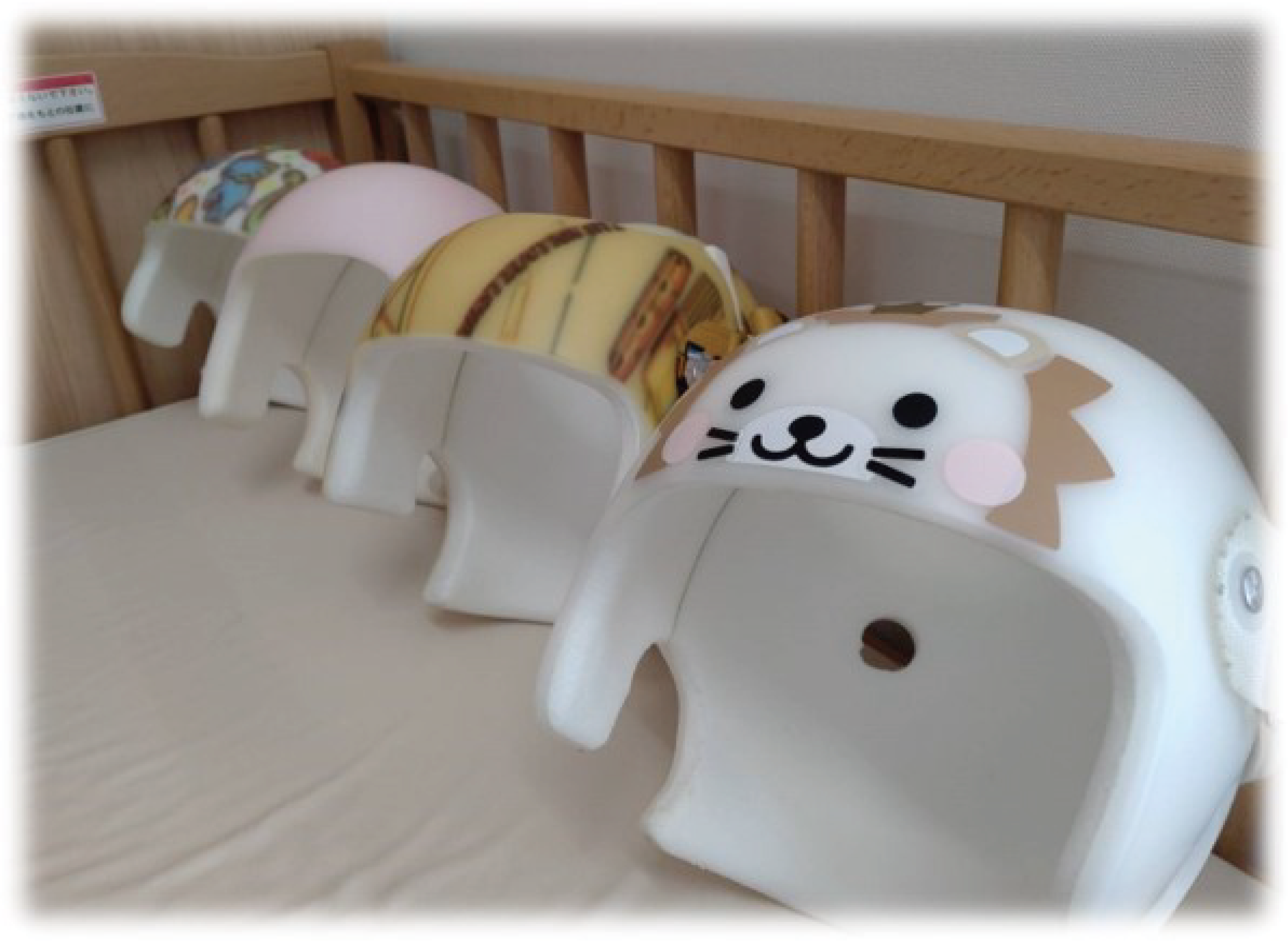
If a moderate or severe degree of deformation is present from the neonatal period to around 2-3 months after birth, the likelihood of natural correction decreases significantly. It is best to consult a specialist outpatient clinic as early as possible, but the appropriate age for starting corrective treatment with a cranial orthosis is between 4 and 18 months of age. The specific timing may vary depending on the type of orthosis, so the specialist will determine the appropriate time to start treatment after conducting an examination.
I heard that there are various diseases that can cause cranial deformation. Please tell me about representative diseases.
Hydrocephalus, craniosynostosis, and syndromic craniosynostosis (such as Crouzon syndrome, Apert syndrome, Pfeiffer syndrome, etc.) are representative diseases that cause cranial deformities. The causes of hydrocephalus include arachnoid cysts, intracranial hemorrhage during childbirth (subependymal hemorrhage, subdural hematoma, etc.), abnormalities in cerebrospinal fluid absorption following meningitis, and genetic or hormonal abnormalities.
I was told that my baby has “torticollis”. I saw on the internet that “torticollis” can cause “positional plagiocephaly”. Is that true?
Both can potentially cause the other. Torticollis can lead to positional plagiocephaly, and positional plagiocephaly is also known to induce torticollis.
When did the helmet treatment for cranial deformities historically begin?
In 1979, Clarren et al. reported cases of infants under one year old with non-synostotic cranial deformities that were successfully corrected using cranial orthotic devices, including severe cases. Since then, numerous studies have emphasized the importance of early intervention for cranial deformities, and the significance of early intervention for cranial deformities is now widely recognized.

1)S.K. Clarren et al. Helmet treatment for plagiocephaly and congenital muscular torticollis J Pediatr (1979)
” I was diagnosed with ‘positional plagiocephaly.’ What does ‘positional’ mean?”
The term “positional” is derived from the literal translation of the English phrase “Positional skull deformity.” Positional plagiocephaly can be improved through changes in the baby’s position or sleep position, particularly in infants who have not yet developed neck control. However, once infants are able to move their heads freely, the effectiveness of frequent position changes or stretches may diminish. The word “positional” can be somewhat misleading and may lead to a mistaken interpretation that parental inattention is the cause of the deformation.
Please tell me what is important in screening for cranial deformation.
The most important opportunity for screening cranial deformities is during the one-month infant check-up. If you feel any discomfort about the shape of your baby’s head, please feel free to consult with us at any time. We understand that you may feel anxious, but some parents have been reassured by the specialist’s examination and explanation, so please do not hesitate to consult with us about any concerns you may have.
How is cranial deformation measured?

Various indices such as Cranial Asymmetry, Cranial Vault Asymmetry Index, and Cephalic Index are commonly used to measure cranial deformation. The index used depends on the type of cranial deformation, so it is recommended to consult a specialist clinic for more information.
My baby has been diagnosed with craniosynostosis. What kind of disease is it?
Craniosynostosis is a condition in which one or more of the sutures of the skull fuse prematurely. It is also referred to as craniofacial synostosis or synostosis syndrome. About 4-10 out of 10,000 births are affected by craniosynostosis. The cause of this condition is not fully understood, but it is believed that intrauterine constraint is one of the factors.1) Genetic abnormalities may also be involved. In 2022, a Dutch study identified the FUZZY gene as a likely candidate for the causative gene of craniosynostosis, but further research is needed.2)
Craniosynostosis can lead to abnormal brain development due to the early fusion of the skull. This can result in symptoms such as increased intracranial pressure and skull deformities, such as scaphocephaly, trigonocephaly, or plagiocephaly, depending on which suture is affected.1)Website: Japan Society of Plastic and Reconstructive Surgery
2)Barrell, W.B., Adel Al-Lami, H., Goos, J.A.C. et al. Identification of a novel variant of the ciliopathic gene FUZZY associated with craniosynostosis. Eur J Hum Genet 30, 282–290 (2022). https://doi.org/10.1038/s41431-021-00988-6
When I look on the internet, I come across the term ‘anterior fontanelle.’ What is it?
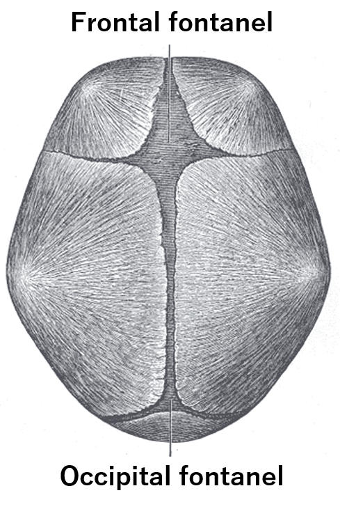
The “anterior fontanelle” is a diamond-shaped gap in the bones located at the front of the skull. The size of the anterior fontanelle is approximately 5 cm in infants from 0 to 6 months of age, 4 cm from 6 to 9 months, and 3 cm in infants between 9 and 12 months old. It usually closes around 18 months of age (between 7 to 15 months after birth).

In conditions such as intracranial hypertension, the tension of the anterior fontanelle can become high, and other suture lines may also separate. During pediatric health check-ups, the anterior fontanelle, posterior fontanelle, and the palpation of the skull sutures are examined.
What are the risk factors for cranial deformation before birth?
Premature birth, hypotonic muscle disorders, congenital torticollis, intrauterine constraint (such as multiple pregnancies, oligohydramnios, etc.) are known to be risk factors for cranial deformation before birth.
1) Littlefield TR, Kelly KM, Pomatto JK, Beals SP. Multiple-birth infants at higher risk for development of deformational plagiocephaly. Pediatrics 1999;103:565-9.
I heard that premature infants are more likely to develop cranial deformation. Is this true?
Premature infants are known to be a risk factor for cranial deformation.1)
1)Littlefield TR, Kelly KM, Pomatto JK, Beals SP. Multiple-birth infants at higher risk for development of deformational plagiocephaly. Pediatrics 1999;103:565-9.
Tell me the causes of cranial deformation.
First, it is necessary to consider the causes of cranial deformation before and after birth. Pre-birth risk factors include abnormal fetal position (pelvic or transverse), multiple pregnancies (twins, triplets, etc.), and abnormalities in uterine shape. The baby’s head is very fragile, and the conditions during delivery have a significant impact. It is known that physical external forces on the fetus in the uterus can cause cranial deformation even before birth.
Post-birth risk factors include premature birth, being a first-born, sleeping in a supine position, and being small-for-date. The frequency of cranial deformation is higher in cases with motor impairments such as cerebral palsy and muscular torticollis, due to difficulties with head flexion, extension, and rotation.What are the types of cranial deformation?
Cranial deformation is classified into categories such as plagiocephaly, brachycephaly, and scaphocephaly. Plagiocephaly is the most common type. It is characterized by the movement of the ear on the same side as the deformation forward, the protrusion of the same side of the forehead, and flattening of the back of the head, all resulting from positional cranial deformation. It is important to differentiate craniosynostosis, a condition that causes cranial deformation, from positional cranial deformation. Craniosynostosis is a congenital abnormality and should be considered as an entirely different disease concept from positional cranial deformation.

1)DEFORMATIONAL PLAGIOCEPHALY: PREVALENCE, QUANTIFICATION AND PREVENTION OF ACQUIRED CRANIAL ASYMMETRY IN INFANTS, Henri Aarnivala, May 2017
How does cranial deformation change depending on the age of the infant?
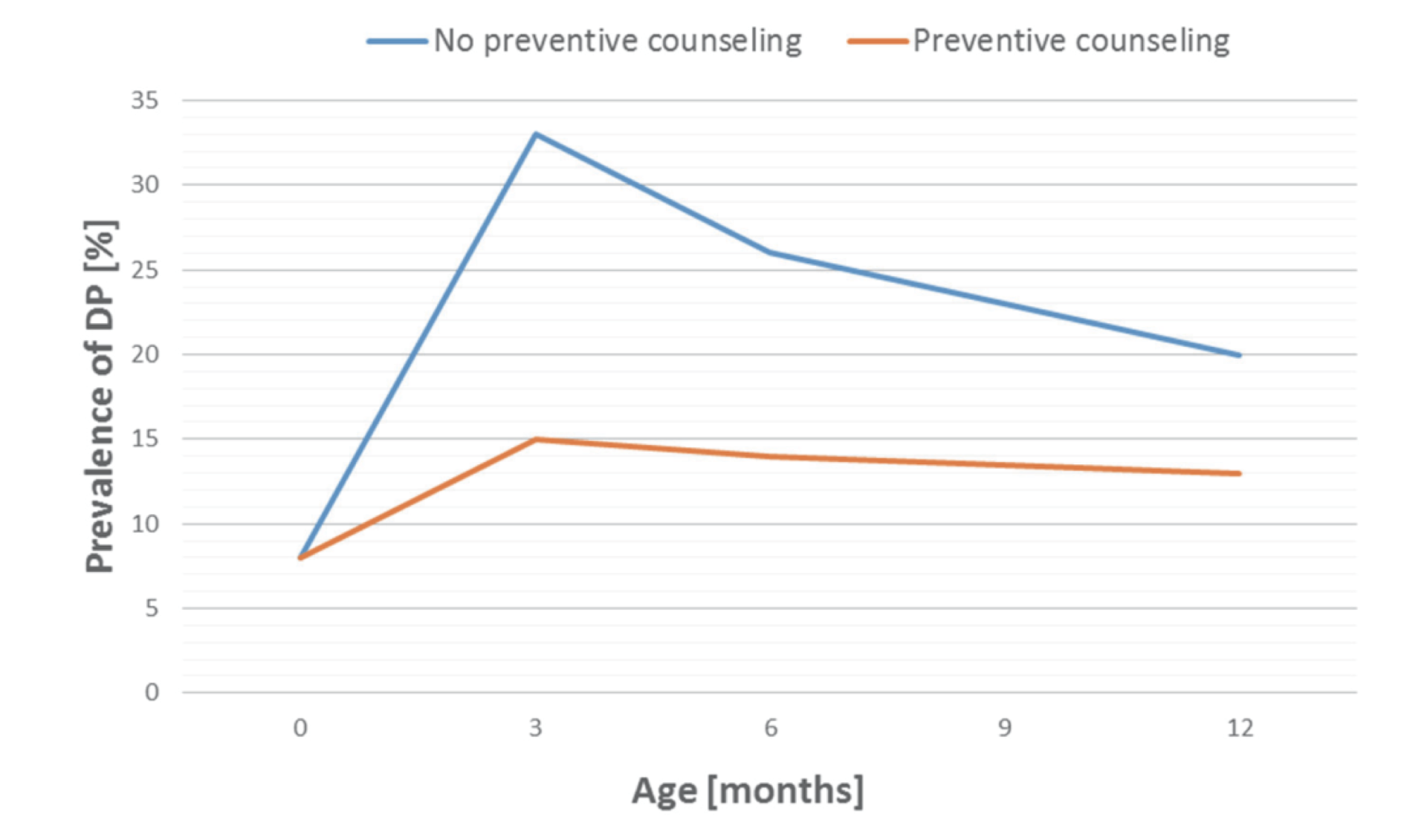
According to data from 2017, the incidence of cranial deformation is reported to be highest around three months of age. It can be confirmed from the table below that cranial deformation has decreased with appropriate intervention (Preventive counselling group).1)
It has been found that positional cranial deformation improves after the infant achieves consistent neck control.

1)DEFORMATIONAL PLAGIOCEPHALY: PREVALENCE, QUANTIFICATION AND PREVENTION OF ACQUIRED CRANIAL ASYMMETRY IN INFANTS, Henri Aarnivala, May 2017
Why is supine sleeping recommended?
As Sudden Infant Death Syndrome (SIDS) became a social issue, the risk of “prone sleeping” became widely known. As a result, “supine sleeping” was recommended, and this is how it came to be promoted.
Is there any reason why head deformity has recently gained attention?
In 1992, the American Academy of Pediatrics (AAP) launched a major campaign called “Back to Sleep,” which aimed to reduce the incidence of SIDS (Sudden Infant Death Syndrome), a social problem at the time, by placing infants in a supine position when sleeping. 1)
Thanks to this campaign, a decrease in the mortality rate was observed between 1992 and 2000. However, on the other hand, flattening of the back of the head has become problematic.2)
Since 1992, the incidence rate of positional skull deformity has been reported to be four times higher. In the United States, it has been reported that one out of every 60 babies has head deformity.1. American Academy of Pediatrics AAP Task Force on Infant Positioning and SIDS: Positioning and SIDS. Pediatrics 1992;89(6 Pt 1):1120-6.
2. Diagnosis and Management of Positional Head Deformity, Wendy S.Biggs, M.D.,University of Michigan Medical School, Ann Arbor, Michigan; American Family Physician, VOLUME 67, NUMBER 9 / MAY 1, 2003
What is the frequency of head deformity?
In early infancy, it has been reported to be observed in approximately 40% of babies, but as there is no unified definition of head deformity, the reports vary widely depending on the paper.
Is there another term used to refer to ‘head deformity’?
The name for head deformity varies depending on the type of deformity. It is referred to by various names, such as positional skull deformity, positional head deformity, oblique head, plagiocephaly, and others. The common name “misshapen head” is also frequently used. Because the causes of head deformity include a variety of conditions, there are multiple names in use to describe it.

What is the definition of head deformity?
The term “head deformity” is widely used, but there is currently no unified definition. Overseas, there is a common name called “baby misshapen head,” but the group of conditions included there is diverse.
I heard that the bones in the skull are a combination of several types of bones. Can you explain in detail?
The skull is composed of eight bones that form a dome surrounding the brain stem during infancy. In adults, the skull bones are all connected to each other by nearly immovable joints called sutures. However, in infants, fibrous sutures are formed but not yet fused. This fusion process typically occurs around the age of 2-3 years.
The unfused bones allow for rapid growth of the brain to occur. Brain growth begins during fetal development, and the brain weighs about 300 grams at birth. By six months of age, it has doubled in weight, and by two years old, it has tripled. It is known that by the ages of 4-6 years old, the brain approaches about 95% of the weight of an adult brain (1200-1500 grams).
ダーマペンについて
施術をお受けできない方
妊娠中・授乳中・膿疱性酒さ・滲出を伴う重度の尋常性ざ瘡・いぼ・ヘルペス・強皮症・日光性角化症・皮膚癌・血友病・鉄アレルギー
※使用する消毒、麻酔や薬剤にアレルギーのある方は事前にお申し出ください。
※その他、医師の判断により施術をお受けできない場合がございます。
ご相談ください。治療後の注意点
治療後12時間は皮膚感染予防および皮膚に導入した薬剤の効果を妨げないようメイクや洗顔を行わず、施術部位は可能な限り清潔にして下さい。
施術当日は、市販の日焼け止めは副反応が起こる可能性があるため使用できません。
※当院では専用の日焼け止めを販売しておりますのでご希望の方はご予約時またはご来院時にお申し付け下さい。副作用/ダウンタイムについて
施術の程度により、赤み、内出血、軽度の出血、腫れやヒリつきが生じることがあります。
ほとんどの場合、3日程度で消失致しますが、長い方でも 1 週間程度となります。
※当院では施術後、鎮静・保湿パックを致します。
プラセンタ注射について
注射を受けた当日はなにか制限はありますか?
当日の入浴なども特に問題ありません。注射部位に違和感、軽度の痛みを感じる場合はありますが、食事や飲酒への制限はありません。現在内服中の薬や既往歴で不安があれば医師にご相談下さい。
保険適応はありますか?
プラセンタ注射剤は更年期障害・乳汁分泌不全・肝機能障害に対し保険適応が認められています。しかし美容を目的としたプラセンタ注射は残念ながら、保険適応外使用となります。当院は自費診療クリニックであり保険適応外となりますので、その点ご理解いただければ幸いです。
FGFやEGFがプラセンタに含まれていると聞きました。どういう効果があるのですか?
プラセンタ抽出注射にはコラーゲンやエラスチンを産生する線維芽細胞を増殖させる線維芽細胞成長因子(FGF)、母体内で胎児の皮膚形成を促す上皮細胞成長因子(EGF)などが含まれていることが知られています。この他にも神経細胞増殖因子(NGF)・コロニー形成刺激因子(CSF)・インシュリン様成長因子(IGF)、その他様々なインターロイキン(Ⅰ~Ⅳ)など、プラセンタに含まれる成長因子は多岐に渡り存在しています。
プラセンタ注射で感染症のリスクはありますか?
各種肝炎ウイルス・HIV・HTLV-1・HPVなどに対し血清学的検査が行われ、感染のリスクはほぼ0に等しくなっています。ただし、このような万全の安全体制の下精製・製造されてはいますが、生物由来製剤であることからどうしても未知のウイルス等に由来する感染症などを理論上完全に否定することは出来ません。まだ見つかっていないウイルス・変性タンパクによる疾病・変異型クロイツフェルト・ヤコブ病(狂牛病の原因となる変異タンパク質)の伝播リスクについては完全には排除できないのが現状です。その為、献血に制限があったり、臓器移植ドナーなどができないなどの制約があります。ただ、国内・海外と50年以上使用されている中でそのような報告はなく、最も安全な生物由来製剤ということができます。
プラセンタの種類を教えてください。
当院ではプラセンタ・エキスの注射剤として『メルスモン』と『ラエンネック』の二種類を採用しています。この二剤はヒト由来の胎盤から精製されたものです。投与方法としては筋肉注射です。注射による投与方法を用いることで、直接作用が期待できることと、代謝を受けずに血中移行することで迅速な作用を期待できるメリットがあります。 現在『プラセンタ由来』を謳うプラセンタ錠剤やプラセンタ配合の美容液・化粧品が一般に販売されています。市販で購入できるものは主に豚や馬などの動物性由来のプラセンタです。内服のものは消化器系で分解され主にアミノ酸として吸収されることになります。効果としては注射剤に遥かに及ばない間接的な摂取方法と考えられます。
プラセンタは男性にも使用してよいのですか?
男性の方も注射可能です。プラセンタ注射により男性ホルモンへの影響などを心配される方もいるかも知れません。「女性化」等の副作用を心配される男性の方もおられますが、その心配はありません。 性別に関係なく、美容・疲労回復・老化防止・肌質の改善などが期待できます。また、男性の更年期障害であるLOH症候群(加齢男性性腺機能低下症候群)などに伴う不調を改善する効果も期待されています。
プラセンタはいつから使用されてきたのですか?
プラセンタの歴史は長く、古くはヒポクラテス(紀元前400年頃)の時代までさかのぼると言われています。古代エジプトの有名な女王クレオパトラも若さを維持するために愛用したと伝えられています。中国では秦の始皇帝の頃から不老長寿の薬として用いられていることが知られており、現在でも『紫河車』という漢方薬名で幅広く活用されました。 近世では、旧ソ連のオデッサ大学の教授であった眼科医の フィラトフ医師(1875-1956)が1920-1930年頃に疾病の治癒に臨床応用したことがわかっています。1956年には注射薬であるメルスモン、ついで1959年にラエンネックが薬事承認を取得し現在に至っています。 現在では医薬品のみならず、健康食品・化粧品にまで使用が広がり、豚や馬のプラセンタが使用されていますが、効果効能については信頼ある参照データが少なく、今後の研究が待たれるところです。
プラセンタには何が含まれますか?
プラセンタの有効成分には、growth factor(成長因子:EGFやFGFなどの成長因子が含まれます)やサイトカイン(インターロイキンなど)、各種必須アミノ酸(リジン、アラニン、アスパラギン酸、ロイシン、グルタミン酸、グリシン、バリン、セリン、チロシン、 フェニルアラニン、スレオニン、アルギニン、プロリン、イソロイシン、メチオニン、ヒスチジン)、酵素、ミネラル、ビタミンなど多く含まれています。
白玉点滴について
妊娠中です。点滴は可能でしょうか?
妊娠中でも点滴は可能です。初回についてはかかりつけの産科医師ともご相談いただくことがありますので、ご了承下さい。
グルタチオンのサプリ・経口薬の効果はあるのでしょうか?
消化器系を介しての吸収となりますので非効率的な摂取方法となります。体内に一度入っても吸収までに時間がかかることが知られており、点滴がその点でも優れていると考えられています。
複数の注射剤を混合して使用すると聞きました。問題はないのでしょうか?
問題ありません。当院では薬剤師と相談の上、最も安全・安心できる点滴組成としています。安心して三鷹ヒロクリニックで白玉点滴を受けていただけます。
にんにく注射について
『にんにく注射』をするとにんにくの臭いが体からするようになるのでしょうか?
もちろんそんなことはありません。注射中は静脈から吸収されたアリナミンの刺激が嗅覚を刺激しアリナミンの臭いを感じることがあります。しかし、にんにくの臭いが周囲に広がるわけではありませんので、安心して下さい。
にんにく注射に興味があります。どのような時に注射を受ければよいのでしょうか?
にんにく注射に含まれる成分は以下のような効果があります。
・疲労回復
肉体疲労時の栄養補給に有効なビタミンであるビタミンB群とビタミンCが含まれていることから、疲労回復には有効な注射となります。 にんにく注射は即効性がある点滴です。「疲労が抜けない」「体調不調」「今は頑張らないといけない」と思ったときに点滴をしていただければよいです。 頻度ですが、週1~2回で行っていただくのがよいです。皆様の体調に合わせて点滴をシていただければ良いかなと思いますので、スタッフにご相談下さい。ビタミンの役割について教えてください。
ビタミンB1は別名チアミンと呼ばれます。体内のエネルギー活動にはブドウ糖は必須です。このビタミンB1はブドウ糖をエネルギー変換するためには必須となる重要なビタミンです。また、粘膜再生・皮膚保護など多くの作用があることから、定期的・継続的に摂取することが進められています。
その他ビタミン群には
・赤血球の製造
・DNAの合成をサポート
・神経機能の正常化
・神経再生
など多くの役割を果たす健康な体を維持するための重要な成分です。
ダイエット点滴について
点滴を受けられないことはありますか?
α-リポ酸が血糖を下げる方向に働くことが知られています。血糖異常、特に糖尿病・内服加療中の方は治療を受けられない場合があります。カウンセリング・診察時にもお話させていただきますので、担当医師にご相談下さい。
副作用などありますか?
投与前後で気分不良・眩暈・嘔吐感・発赤などがまれに起こることがあります。また治療を続けた結果としての長期的な副作用は報告されていません。詳しくは担当医師にご相談下さい。
どれぐらい続ければいいでしょうか?
治療効果を十分に引き出すためには点滴を一ヶ月2〜4回行い、運動・食事療法を併用することが有効だと言われています。
点滴を受ければ必ず痩せますか?
脂肪燃焼を促進する点滴ではありますが、単独では効果はあまり望めません。適切な運動と食事療法を併用することがより効果的となります。
すぐに効果がでますか?
ゆるやかな体質改善を目指し健康的に体重減少を目標とすることから、注射後すぐに劇的な効果が現れるわけではありません。しかし、数ヶ月経過して効果を実感している方が多いです。
点滴時間はどれぐらいですか?
カウンセリングから点滴終了までおおよそ20分から40分ほどです。
Reservation ご予約はこちらから
Web予約限定5大特典
- 1.お肌のカウンセリング 完全無料
- 2.お肌トラブル時の診察代 完全無料
- 3.前日までの予約キャンセル料 完全無料
- 4.他部位へのテスト照射 完全無料
- 5.万一の照射漏れの際の追加照射 完全無料
- ※5大特典には条件がございます。
Useful Information お役立ち情報
View allPopular Articles よく読まれているコンテンツ

医療脱毛の施術を受ける前日・当日の生活で意識したい10の注意点
2022.05.23
脱毛メカニズム
顔にニキビができても脱毛できる?OK・NGのケースをチェック
2022.11.11
脱毛メカニズム
ニキビ治療と漢方薬 ☆桂枝茯苓丸加薏苡仁・清上防風湯・十味敗毒湯☆
2023.06.18
医療情報トピック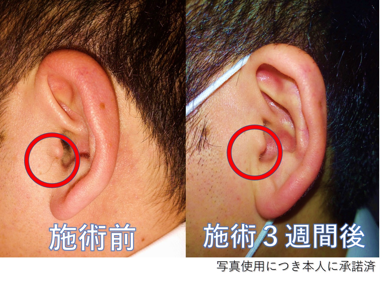
耳毛(医療脱毛)
2022.02.11
脱毛トピック


